See a clinical case
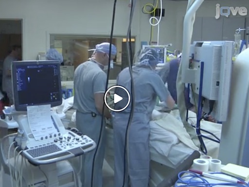
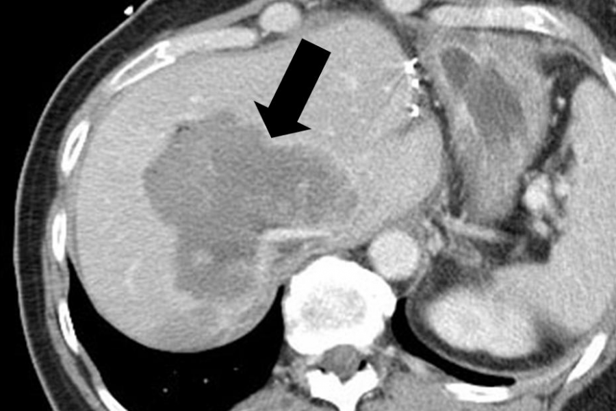
Tumor ablation is a minimally invasive therapy used to treat cancers with well-defined tumors, especially those in the liver, kidney, lung and bone. Medical imaging is used to plan, guide and monitor most treatments. Most patients can be treated and released in 24 h with minimal pain and few complications. A typical case is presented in this video.
Read about the technology
During ablation, tumor cells are targeted and killed by applying heat, cold, chemicals, electric fields or intense ultrasound. Most devices are needle-like and be inserted directly into the tumor to create highly focal tissue destruction. Some are completely non-invasive. The pros and cons of available technologies are highlighted in this article.
Research Topics
Antenna Design
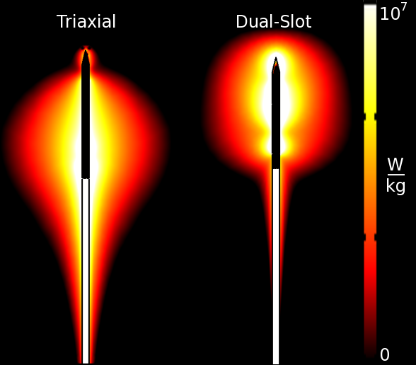
Microwave ablation antenna design determines how efficiently and in what geometric pattern energy is delivered. We aim to improve the efficiency and heating geometry of our devices to address a range of clinical indications.
Techniques
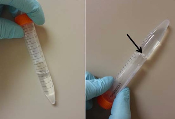
Hydrodissection is injecting a fluid or gel material to physically separate and protect non-target tissues, and can be used to facilitate ablation in difficult anatomic locations.
Computer Simulation
Simulations allow us to predict how devices will interact with tissue. As numerical techniques improve, we can perform experiments "in silico" to study the relevant biophysics and optimize treatments.
Clinical Translation
Our research has a singular focus: impact patient care. We move quickly to translate our developments into clinical practice, whether by adapting clinical workflow or through the commercialization of novel devices.
Imaging Feedback
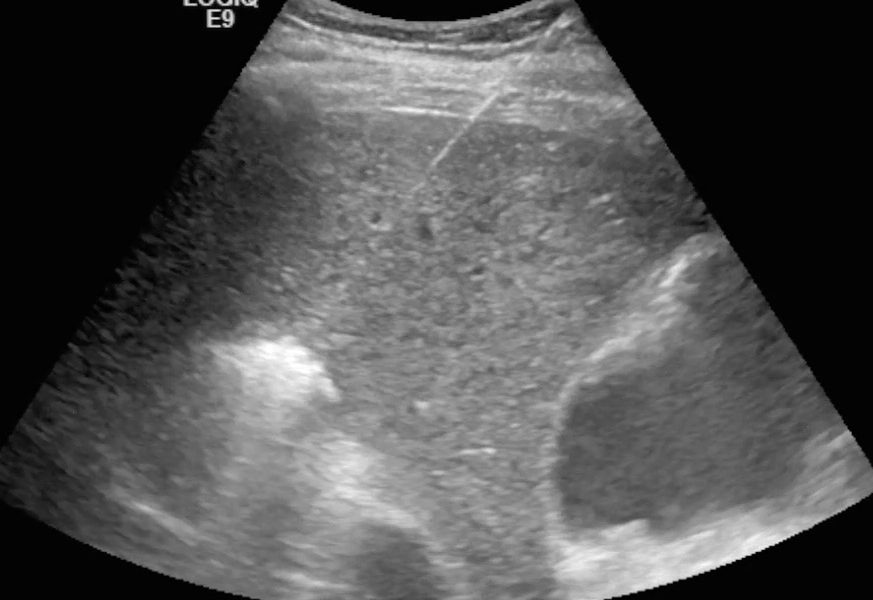
Medical imaging such as ultrasound, CT and MRI are used to guide applicator placement, monitor the treatment, and post-procedure follow-up. We aim to improve each phase of the treatment. Projects involve collaborations with various resources on campus, including Radiology, Medical Physics, and the Center for High Throughput Computing.
